Es tut uns leid: Aufgrund des Auflösungsbeschlusses wurden alle Vereinsaktivitäten eingestellt.
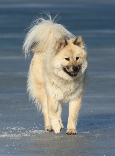
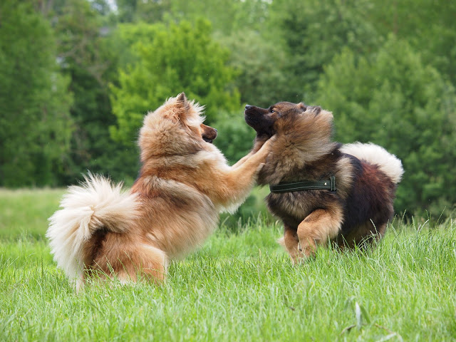

Eurasier - Rassehund - ein klasse Hund
Es tut uns leid: Aufgrund des Auflösungsbeschlusses wurden alle Vereinsaktivitäten eingestellt.
© 2024 linkXpert Schaetz